Navicular Syndrome Explained
Oct 07, 2015
Published in Saddle Up Magazine September 2015
Most horse owners cringe at hearing the words Navicular Syndrome. In the past it has often meant expensive corrective shoeing and just trying to keep your horse sound for one more season, inevitably resulting in putting them down. Unfortunately Navicular Syndrome is one of the hardest subjects for a farrier/trimmer to research. It seems that every old text contradicts the next, and every person you talk to has a different understanding of the condition. The good news is that tons of new research is being done, and hopefully the equine world will soon have a better understanding that the best way to treat Navicular Syndrome is to prevent it in the first place, and that it is easy to do so.
One of the doctors at the forefront of Navicular research is Dr. Robert Bowker of Michigan State University. I had the pleasure of meeting Dr. Bowker at the 2015 Equine Education Summit hosted by Horse Council BC this past spring. Dr. Bowker states that he has identified a heel first landing as the most important element of hoof function and more importantly hoof development. He has determined that as the hoof impacts the groundheel first, the hoof expands both laterally and from the back to the front, and the concave sole descends lower to the ground, thus dramatically increasing the volume of the hoof capsule. This sudden increase in volume creates a vacuum, which pulls blood into the hoof capsule. This pull of blood not only nourishes the living tissues in the hoof, but acts as a very important hydraulic shock absorber.

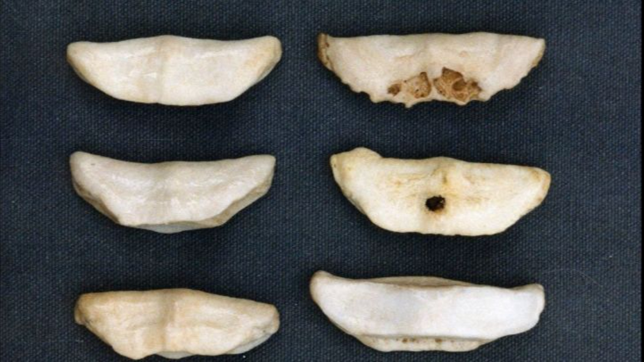
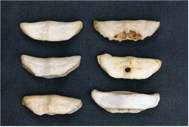
Healthy navicular bones and navicular bones with permanent damage
It has been documented over the years that some horses with navicular bone changes are perfectly sound, while others without bone damage can show severe lameness in the rear of the hoof. This is confusing though as it has long been taught in the veterinary and farrier communities that the bone damage happened first and the pain associated with Navicular Syndrome was caused by the deep digital flexor tendon sliding over the rough surface of the damaged navicular bone.
Dr. James R. Rooney of the American College of Veterinarian Pathologists specializes in post mortem studies of horses. In thousands of dead horses he has examined, Dr. Rooney found that the fibrocartilages surrounding the flexor tendon and navicular bone were always damaged if bone deterioration was present. He has not found one single case where the bone was damaged and the fibrocartilage was not. He has however found cases where the bone was not yet deteriorated and yet the fibrocartilage had started to break down. He has learned that the order in which damage occurs is: first the fibrocartilages surrounding the navicular bone, second the fibrocartilages surrounding the deep digital flexor tendon, third the flexor tendon itself, and finally the navicular bone itself is damaged by the rough surface of the damaged flexor tendon. Simulating a toe first landing with cadaver horse legs in test machines, Dr. Rooney was able to simulate this exact process of deterioration, proving the order in which tissues were damaged leading to Navicular Syndrome.
The main structure in the front half of the hoof is the coffin bone. The sole and hoof wall are attached to it via the lamina. The digital cushion and the lateral cartilages form the rear half of the hoof. It is the rear half of the hoof that is responsible for dissipating the impact energy of movement. The front of the hoof has no impact absorbing structures, they are all fairly rigid. The digital cushion at birth is made up of primarily fat and is filled with nerves. As the foal moves the pressure and release of the frog causes fibrocartilage to grow from the front of the digital cushion and spread toward the back. By the time the horse has grown to an adult the digital cushion should have transformed into a mass of fibrocartilage. This fibrocartilage is responsible for protecting and cushioning the nerves as well as dissipating the energy of a heel first landing. The lateral cartilages at birth are tiny, less than 1/16th of an inch thick, and don’t extend to the underside of the frog and digital cushion yet. As the foal grows, with movement, flexion, and the expansion and contraction of the hoof mechanism the lateral cartilages grow. They should eventually extend to create a floor underneath the frog and digital cushion and should have developed to about an inch thick.
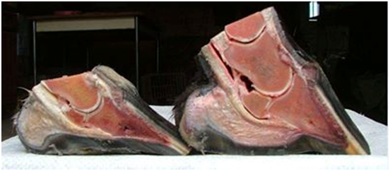
So why are most horse’s uncomfortable landing heel first? Because in domestication we tend to keep our foals on soft ground. Deeply bedding the stalls that restricts their movement, and keeping them on soft terrain when they are turned out. The soft ground inhibits the flexion, expansion and contract and negates the hoof mechanism as it was designed to work. This results in very commonly, adult horses with lateral cartilages as thin as 1/8th of an inch thick instead of the inch they should be, and with digital cushions that are underdeveloped, thin and weak.

On the left an atrophied and weak digital cushion, on the right a healthy digital cushion
Dr. Bowker has also found that bone loss associated with Navicular Syndrome can also be attributed to a lack of natural pressure in the navicular region of the hoof. He specifically blames peripheral loading i.e. shoeing the hoof to remove sole pressure or allowing the hoof wall to grow too long so that the sole, frog, and bars of the hoof cannot share in the weight baring pressures of movement as they were designed.
When we learn the science behind Navicular Syndrome, and when this information becomes mainstream, only then can we start to prevent these changes from happening. While we cannot heal the bone deterioration once it has happened, we can bring strength back to the digital cushion and lateral cartilages. We must first bring them back into work, by removing the peripheral loading devices, keeping a low heel and allowing the digital cushion to strengthen again. The digital cushion is filled with myoxoid tissue which is similar to stem cell tissue and Dr. Deborah Taylor of Auburn University has published that the digital cushion can regenerate if given the opportunity. And as discussed previously, horses with bone deterioration to the navicular bone can be made comfortable if the rest of the hoof is allowed to strengthen to support it.
If your horse is suffering from Navicular Syndrome or you want to learn more I would direct you to study the research of Dr. Robert Bowker, Dr. James R. Rooney and Dr. Deborah Taylor. They are leading the research right now, and are coming up with amazing information that is helping horses that would have previously been put down.
